
Post by joeysgreen on Mar 13, 2007 16:03:55 GMT -5
Hey guys, I did another necropsy the other day. Thanks Rhiannon for the body, and for allowing me to share these photos. A bit of history, this was a monkey tail skink that died 5 days after bringing him into a new home. He was very dehydrated upon arrival.
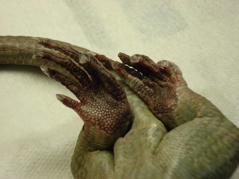
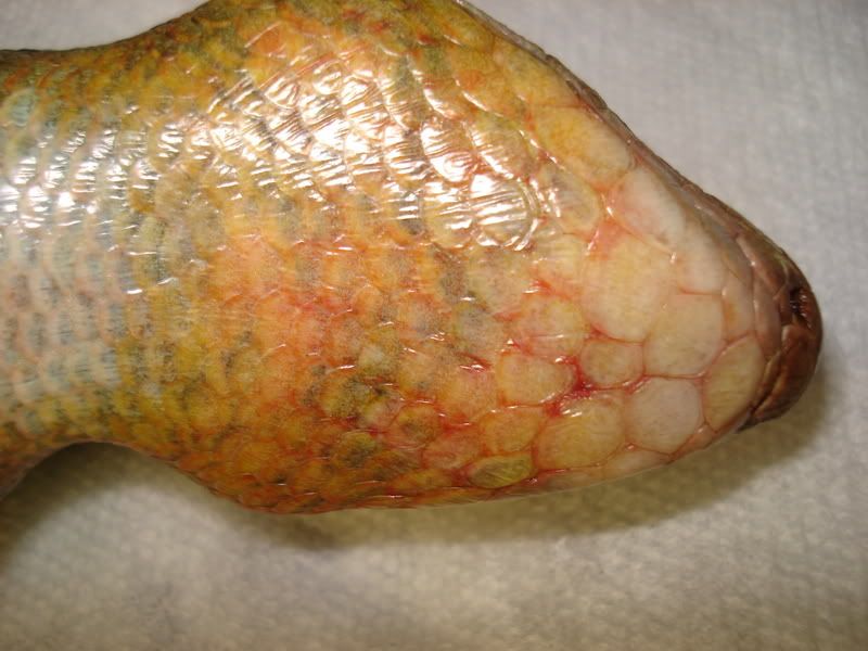
Now with a necropsy (non-human autopsy), much can be learned before even grabbing for the scalpal. This animal was very skinny; border-line emaciated. Dehydration is tough to identify post mortem due to natural changes that occur at this time. I don't doubt that this animal was quite dehydrated from Rhiannon's description. After the immediate impression, a closer look at the extremities and skin found what looked like burns or other dermal lesions. Erythemia was present on the throat, neck, hands and feet. The hind limbs were the worst.
Internally, there wasn't much to be found. Everything looked pretty normal. The stomach was empty, however the intestines were packed solid with bright green chyme/stool. I don't think this was an impaction, however gut stasis can happen with severe dehydration. I don't know what killed this animal, however whatever the instigator, this animal was losing weight, dehydrated, and septic. I would highly consider an untreated thermal burn as a cause.
For the educational half of the necrospy, I focused on the head anatomy.
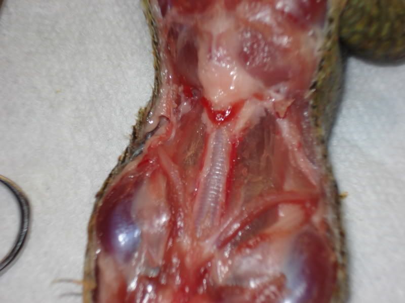
The red "V" is the thyroid gland. The ribbed tube running underneath it is the trachea. Underneath is a membrane covering the throat. Sorry about the crappy photo, but it's the only one I had that showed off the thyroid. Something you don't see every day.
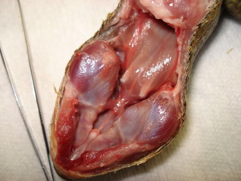
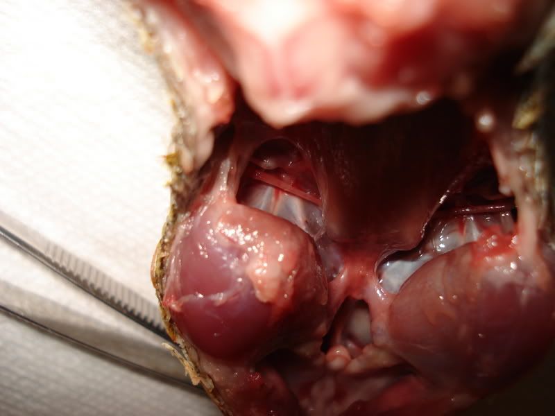
Further dissection shows the two prominent mastication muscle groups. The grooves running downwards in the middle are the nares/choana area. Unlike humans and most mammals, there is no hard palate that divides the mouth and nasal cavity. The enlongate, whitish tissue is the Jacobson's organ. Most interestingly was the grooves up and around the aforementioned muscles that were direct openings to the middle ears. The next picture shows this in more detail.
Here you can see the bones responsible for channeling vibrations to the ear's nervous centre (inner ear). The tympanic membrane isn't visible, but ajoins the long thin bone on the left hand or lateral side. The openess surprised me and there is a chance that a membrane had descicated post mortem, however I saw no evidence of such.
Anyhow, I learned alot, and I hope you enjoyed this post.
Ian
Now with a necropsy (non-human autopsy), much can be learned before even grabbing for the scalpal. This animal was very skinny; border-line emaciated. Dehydration is tough to identify post mortem due to natural changes that occur at this time. I don't doubt that this animal was quite dehydrated from Rhiannon's description. After the immediate impression, a closer look at the extremities and skin found what looked like burns or other dermal lesions. Erythemia was present on the throat, neck, hands and feet. The hind limbs were the worst.
Internally, there wasn't much to be found. Everything looked pretty normal. The stomach was empty, however the intestines were packed solid with bright green chyme/stool. I don't think this was an impaction, however gut stasis can happen with severe dehydration. I don't know what killed this animal, however whatever the instigator, this animal was losing weight, dehydrated, and septic. I would highly consider an untreated thermal burn as a cause.
For the educational half of the necrospy, I focused on the head anatomy.
The red "V" is the thyroid gland. The ribbed tube running underneath it is the trachea. Underneath is a membrane covering the throat. Sorry about the crappy photo, but it's the only one I had that showed off the thyroid. Something you don't see every day.
Further dissection shows the two prominent mastication muscle groups. The grooves running downwards in the middle are the nares/choana area. Unlike humans and most mammals, there is no hard palate that divides the mouth and nasal cavity. The enlongate, whitish tissue is the Jacobson's organ. Most interestingly was the grooves up and around the aforementioned muscles that were direct openings to the middle ears. The next picture shows this in more detail.
Here you can see the bones responsible for channeling vibrations to the ear's nervous centre (inner ear). The tympanic membrane isn't visible, but ajoins the long thin bone on the left hand or lateral side. The openess surprised me and there is a chance that a membrane had descicated post mortem, however I saw no evidence of such.
Anyhow, I learned alot, and I hope you enjoyed this post.

Ian
